このアプリケーションノートでは、ナノ毒性学および生態毒性学の研究の前段階として、ナノサイト装置シリーズでナノ粒子トラッキング解析(NTA)を使用し、テスト媒体に含まれるナノ粒子の状態、特に粒子径、粒度分布、および濃度を特定する方法について説明します。
操作したナノ粒子(NP)の開発と使用が急速に成長しており、そのリスクの評価と管理の方法論よりも先行し続けています。 ナノスケールの粒子径から得られる新規特性は、ナノ材料の性能を裏付ける基礎です。 バルク形態の製品を取り巻く毒性学が十分理解されている場合でも、ナノスケールの同一物質の特性の自動的な「読み取り」は自動仮定とはかけ離れています。ナノスケールの材料は、本質的に新規材料です。 これらの材料の長期にわたる潜在的な毒性作用、および環境に対する潜在的な影響がよく理解されていないという認識が高まっています。 この結果、操作されたナノ材料の潜在的な毒性作用に関する強い興味と研究活動があり、それに伴って既存の製品および調合内に存在するナノスケール材料の割合の重要性が見直されています。
ナノ毒性学の研究を開始する前に、使用するナノ粒子の状態、特に適切なテスト媒体に含まれるナノ粒子の粒子径、粒度分布、および濃度を知ることが重要です。
粒子径により、拡散速度、生物学的障壁による侵入または排除、および粒子間の相互作用が決まります。
このアプリケーションノートでは、生体関連液体中での金ナノ粒子およびその凝集体の特性評価におけるナノ粒子トラッキング解析(NTA)の有用性について説明します。 NTAは従来の光拡散法を補完し、懸濁液中のナノ粒子の径の測定と計数が粒子単位で可能なので、高い分解能で凝集を理解できます。 この研究では、標準分散媒(クエン酸緩衝液)と希釈血漿(タンパク質含有)の場合を比較して、金ナノ粒子の径の変化を調べました(NIST、2007)。
健康なドナーから血液を採取しました。 試験管を800 RCFで5分間遠心分離し、赤血球と白血球をペレット状に分離しました。 上澄み(血漿)をラベル付きの試験管に移し、-80 ˚Cで保管しました。 解凍時に血漿を16.1 kRCFで3分間遠心分離し、さらに赤血球と白血球の含有量を低減しました。 ペレットを乱さないように注意して、この上澄みを新しい容器に移しました。
直径60 nmのNISTの金標準ナノ粒子を使用しました(NIST基準試料8013)。 これらの試料の保管、前処理、および使用は、NISTの関連調査報告書(2007)に従って行いました。 pH 7.19の標準クエン酸緩衝液を使用して、金を約108粒子/mLの濃度に希釈しました。 血漿中に拡散するために、ヒト血漿とクエン酸緩衝液の割合が1:106になるようにヒト血漿を希釈し、金ナノ粒子を含む10 µLのクエン酸緩衝液を790 µLの希釈血漿で希釈しました。
ナノサイトLM10でナノ粒子トラッキング解析を実行しました。 試料の前処理と測定はすべて、アイルランドのダブリン大学で実施しました。 統計的な不変性を確保するために、それぞれが166秒のビデオを複数録画し、バッチモードで解析しました。 血漿は天然のイオン媒体であるため、タンパク質凝集が常に疑われます。
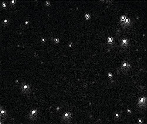
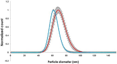
ナノ粒子のビデオを記録し(図1)、クエン酸緩衝液および血漿で希釈した時点で粒子径と濃度の追跡と分析を行い、トラック長で補正しました(図2)。

|

|
クエン酸緩衝液で希釈した金の測定手法は、NISTの基準試料報告書(2007)に詳細に記載されています。
NTAを使用して視覚的に、また濃度測定により、粒子がまだ単分散していることを確認でき、これは懸濁液中のタンパク質がナノ粒子の被膜として機能する(および粒子径の増大は凝集によるものではない)という仮定の根拠になります。
図1および2に記録された粒子径の変化は、ナノ粒子径が約10 nmに増加したことを示します。これは、ナノ粒子を覆う吸着したタンパク質層の厚さ5 nmに相当します。
ヒト血漿のような生体媒体の存在下における単分散金ナノ粒子の粒度分布に有意で測定可能な変化が見られました。 血漿層の厚さの測定にナノ粒子トラッキング解析を用いる適切な方法論をここで説明しました。
クエン酸に分散した粒子についてNTAで測定した広がりと平均値は、NISTの測定に近い値になっています。 血漿の存在下では、粒子径と粒度分布の両方が同様に増加しています。
NTAには絶対濃度を測定する機能があり、NISTの金ナノ粒子の個数濃度を求める目的にも使用できます。 また、NTAは、簡単に区別可能なダイマーなどのナノ粒子凝集体の同定と計数に理想的です。 このため、NTAは、操作した生物学的ナノ材料に関するバイオナノ科学およびバイオナノ相互作用について使用できる一連の既存の手法の中で、(ここで使用したナノ粒子と濃度領域について)有益なツールです。
NTAは、ナノ粒子の粒子径、および同等に重要な項目として濃度に関する情報を提供する解析方法として認識されており、多分散性が高い複雑な試料でも情報提供が可能です(Montes-Burgosら、2010。Lynch、2008。Montes-Burgosら、2007)。 NTAのような方法は、今後、ナノ粒子の環境に対する影響や潜在的な細胞毒性を研究するための多数の手段のうちの1つとして見なすことができます(Bormら、2006。Tenuta、2008。TranおよびAnton、2009。Kuhlbuschら、2010。HassellövおよびKaegi、2009。Stolpeら、2011)。
最近の研究で、NPと環境マトリックスとの相互作用が非常に複雑であり、定量とモデル化の両方が非常に困難であること、しかし定量とモデル化についてNTAが調査と解析のメカニズムを提供可能であることが示されました(Gornatiら、2009。Hartmann、2011。Arvidssonら、2011。Howard、2010。Njugunaら、2011。Tranら、2009)。
主要な海洋無脊椎種の3種の初期発育段階の成長時においてゼロ価鉄ナノ粒子(nZVI)に塗布した被膜の効果の研究で、Kadarら (2012)はNTAを使用して海水に溶解したnZVIを研究し、被膜がナノ金属懸濁液の安定化を促進することを示しました。 また、Kadarは、産業関連で操作されたNTA解析済みの鉄ナノ粒子が海洋微細藻類の培地の成長状態と代謝状態に及ぼす影響も研究し、成長率、粒度分布、脂質像、および細胞の微細構造のその後の変化を説明しました(Kadarら、2012)。
銀粒子固有の毒性と銀イオンの毒性を区別し、クエン酸被膜を持つ銀ナノ粒子とポリビニルピロリドン被膜を持つ銀ナノ粒子の曝露バイオマーカーを開発する目的で、オオミジンコ、淡水甲殻類、および一般的な毒性指標種の15kオリゴヌクレチオドのマイクロアレイを使用して、Poyntonら (2012)は、それらのゲノムレベルでの毒性を研究する前段階として、NTAを使用して銀ナノ粒子の凝集度を測定しました。
トビムシに対する酸化亜鉛ナノ粒子の毒性の研究で、Waalewijn-Koolら (2012)は、分散粒子径の特性のテストに使用する曝露媒体のスパイク方法の違いによって、生物の生殖能力の差がほとんど発生しないことを示し、NTAとTEMの両方で塩化亜鉛の毒性が粒子径に関係しないことを示しました。
細胞レベルで、ヒト末梢白血球に含まれるコバルトナノ粒子の遺伝毒性(Colognatoら、2008)およびマウス線維芽細胞に含まれるコバルトナノ粒子の遺伝毒性(Pontiら、2009)の研究にNTAが有用であることが実証されています。 ナノ粒子がヒト胎盤を通過する能力(Wickら、2009)、および他の生体障壁に対するナノ粒子の影響を研究する方法への取り組み(Linnら、2010)が行われました。ヒト皮膚経由の二酸化ケイ素の輸送(Staroňováら、2012)などがあります。 Similarly, Filon et al. (2012)は無損傷皮膚および損傷皮膚を経由するコバルトナノ粒子のヒト皮膚侵入を報告し、ナノ粒子として塗布したコバルトがインビトロ拡散システム内でヒト皮膚に侵入可能であることを示唆しました。
細胞毒性学的テストの目的で細胞系に導入する前に、ナノ粒子の粒度分布を知っておくことは重要であり、NTAはこの点で有用であることが実証されています(Kendallら、2009。Patelら、2010。Munaro、2010。Karlsson、2010)。 血清(Treuelら、2010)、有機汚染物質(Ben-Mosheら、2009)、ジチオスレイトール(Sauvainら、2008)など、生体由来の様々なマトリックスと各種のナノ粒子との化学的相互作用も研究されました。
肺、肝臓、腎臓、腸、および免疫系を表す各種の株細胞を使用して、コバルトナノ粒子(Co-NP)凝集体の毒性学的な影響が調べられました。 全体的な発見は、コバルトナノ粒子の凝集体の毒性学的な影響は主に凝集したコバルトナノ粒子からのコバルトイオンの溶解に起因するという仮説と一致しました (Limorら、2011)。
Arvidsson R, Molander S, Sanden BA and Hassellov M (2011) Challenges in Exposure Modeling of Nanoparticles in Aquatic Environments, Human and Ecological Risk Assessment: An International Journal, Volume 17, Issue 1, 2011, Pages 245 - 262, DOI: 10.1080/10807039.2011.538639
Ben-Moshe T, Dror I and Berkowitz B (2009) Oxidation of organic pollutants in aqueous solutions by nanosized copper oxide catalysts, Applied Catalysis B: Environmental, Volume 85, Issues 3-4, Pages 207-211
Borm P, Klaessig FC, Landry TD, Moudgil B, Pauluhn J, Thomas K, Trottier R and Wood S (2006) Research Strategies for Safety Evaluation of Nanomaterials, Part V: Role of Dissolution in Biological Fate and Effects of Nanoscale Particles, Toxicological Sciences, 90 (1): 23-32
Colognato R, Bonelli A, Ponti J, Farina M, Bergamaschi E, Sabbioni E and Migliore L (2008) Comparative genotoxicity of cobalt nanoparticles and ions on human peripheral leukocytes in vitro, Mutagenesis Advance Access, published online 2008/05/25. and Mutagenesis 2008 23(5):377-382
Filon FL, Crosera M, Timeus E, Adami G, Bovenzi M, Ponti J, Maina G (2012) Human Skin Penetration of Cobalt Nanoparticles Through Intact and Damaged Skin, Toxicology in vitro, http://dx.doi.org/10.1016/j.tiv.2012.09.007,
Gornati R, Papis E, Di Gioacchino M, Sabbioni E, Dalle-Donne I, Milzani A and Bernardini G (2009) In vivo and in vitro Models for Nanotoxicology Testing, in Nanotoxicity (eds S. C. Sahu and D. A. Casciano), John Wiley & Sons, Ltd, Chichester, UK. DOI: 10.1002/9780470747803.ch15
Hartmann NB (2011) Ecotoxicity of engineered nanoparticles to freshwater organisms, PhD Thesis April 2011, Department of Environmental Engineering, Technical University of Denmark
Hassellöv M and Kaegi R (2009) Analysis and characterization of Manufactured Nanoparticles in Aquatic Environments. In: "Nanoscience and Nanotechnology: Environmental and human health implications." (Eds. Lead J.R. and Smith E.) Wiley Interscience, Chapter 6, p 211-266.
Howard AG (2010) On the challenge of quantifying man-made nanoparticles in the aquatic environment, J. Environmental Monitoring, 12, 135 - 142. DOI: 10.1039/b913681a
Kadar E, Rooks P, Lakey C, Whitea DA (2012) The effect of engineered iron nanoparticles on growth and metabolic status of marine microalgae cultures, Science of The Total Environment, Volume 439, 15 November 2012, Pages 8-17
Karlsson HL (2010) The comet assay in nanotoxicology research, Analytical and Bioanalytical Chemistry DOI: 10.1007/s00216-010-3977-0
Kendall M, Ding P, Kendall K and Clark H (2009) Nanotoxicology of PM: Particle interactions with lung surfactant polymers, IEH (2009) Proceedings of the Annual UK Review Meeting on Outdoor and Indoor Air Pollution Research, 20-21 April 2009 (Web Report W26), Institute of Environment and Health, Cranfield University, UK, available at: http://www.cranfield.ac.uk/health/ieh
Kuhlbusch TAJ, Fissan H and Asbach C (2010) Measurement and Detection of Nanoparticles within the Environment. Nanotechnology. p229-266
Limor H-A, James KC., Rafi K, Patrice NM, Oded M, Ponti J, Romano R, Rossi F, Golla-Schindler U, Sommer D, Uboldi C, Unger R and Villiers C (2011) Predictive toxicology of cobalt nanoparticles and ions: comparative in vitro study of different cellular models using methods of knowledge discovery from data, Toxicol. Sci. (2011) DOI: 10.1093/toxsci/kfr124 First published online: 2011/05/20
Linn M, Loretz B, Philippi C, Vajda V (2010) Optical characterization of nanoparticles, 8th International Conference and Workshop on Biological Barriers - in vitro Tools, Nanotoxicology, and Nanomedicine, 21 March - 1 April 2010, Saarland University, Saarbrücken, Germany
Lynch I (2008), NanoInteract - dispersion,cell culture standards, protocols, NanoImpactNet WP1 Workshop, UCD, Ireland, 20th June 2008.
Montes-Burgos I, Salvati A, Lynch I, Dawson K (2007), Characterization techniques for nanoparticle dispersion, at European Science Foundation (ESF) Research Conference on Probing Interactions between Nanoparticles/Biomaterials and Biological Systems, Sant Feliu de Guixols, Spain, 3 - 8 November 2007
Montes-Burgos I, Walczyk D, Hole P, Smith J, Lynch I and Dawson K (2010) Characterization of Nanoparticle Size and State Prior to Nanotoxicological Studies, Journal of Nanoparticle Research, Volume 12, Number 1 / January, 2010 DOI: 10.1007/s11051-009-9774-z
Munaro B (2010) Mechanistic in vitro tests for genotoxicity and carcinogenicity of heavy metals and their nanoparticles, Dissertation zur Erlangung des akademischen Grades des Doktors der Naturwissenschaften Eingereicht im Fachbereich Biologie an der Universität Konstanz vorgelegt von June 2009 Konstanzer Online-Publikations-System (KOPS) URN: http://nbn-resolving.de/urn:nbn:de:bsz:352-opus-121714
National Institute of Standards & Technology, Report of Investigation, Reference Material 8013, Gold Nanoparticles, Nominal 60 nm Diameter 13/12/2007.
Njuguna J, Sachse S, Silva F, Irfan A, Michałowski S, Pielichowski K, Kazmina O, Ermini V, Zhu H and Blázquez M (2011) Investigations into nanoparticles generated from nanofiller reinforced polymer nanocomposites during structural testing, Safety issues of nanomaterials along their life cycle, Symposium at LEITAT Technological Center, Barcelona (Spain). 4th and 5th May 2011
Patel D, Kell A, Simard B, Xiang B, Lin HY and Tian G (2010) The cell labeling efficacy, cytotoxicity and relaxivity of copper-activated MRI/PET imaging contrast agents, Biomaterials, DOI:10.1016/j.biomaterials.2010.10.013
Ponti J, Sabbioni E, Munaro B, Broggi F, Marmorato P, Franchini F, Colognato R and Rossi F (2009) Genotoxicity and morphological transformation induced by cobalt nanoparticles and cobalt chloride: an in vitro study in Balb/3T3 mouse fibroblasts, Mutagenesis, Jul 2009; DOI:10.1093/mutage/gep027
Poynton HC, Lazorchak JM, Impellitteri CA, Blalock BJ, Rogers K, Allen J, Loguinov AV, Heckman L and Govindasmawy S (2012) Toxicogenomic Responses of Nanotoxicity in Daphnia magna Exposed to Silver Nitrate and Coated Silver Nanoparticles, Environ. Sci. Technol., DOI: 10.1021/es3001618
Sauvain J, Deslarzes S and Riediker M (2008) Nanoparticle reactivity toward dithiothreitol, Nanotoxicology, 2:3, 121 - 129
Staroňová K, Nielsen JB, Roursgaard MJ, Knudsen LE (2012) Transport of SiO2 Nanoparticles through Human Skin, Basic & Clinical Pharmacology & Toxicology, DOI: 10.1111/j.1742-7843.2012.00873.x
Stolpe B, Lead J, Cole P, Kendall M, Kadar E, Poole J, Whitby C, Colbeck I, Fabrega J and Galloway T (2011) Multimethod characterization of manufactured nanoparticles in toxicity studies, 6th International Conference on the Environmental Effects of Nanoparticles and Nanomaterials, N1.7,The Royal Society, London, 19th-21st September, 2011.
Tenuta T (2008) A Systematic Approach to Assessing Potential Environmental Impacts of Nanomaterials: Nanoparticle Synthesis, Characterization and Impact Assessment, , EPA Scholarship & Fellowship Seminar - 13th November 2008, Hilton Kilmainham Hotel, Dublin 8, Ireland
Tran L and Antón JMN (2009) Nanotoxicology And Engineered Nanoparticle Risk Assessment, Seguridad y Medio Ambiente - Nº 114, p1 de 45
Treuel L, Malissek M, Gebauer JS and Zellner R (2010) The Influence of Surface Composition of Nanoparticles on their Interactions with Serum Albumin, Chem Phys Chem, Volume 11, Issue 14, pages 3093-3099
Waalewijn-Kool PL, Ortiz MD and van Gestel CAM (2012) Effect of different spiking procedures on the distribution and toxicity of ZnO nanoparticles in soil, Ecotoxicology. DOI: 10.1007/s10646-012-0914-3Online First™Open Access
Wick P, Malek A, Manser P, Meili D, Maeder-Althaus X, Diener L, Diener P-A, Zisch A, Krug H F. and von Mandach U (2009) Barrier Capacity of Human Placenta for Nanosized Materials, Environmental Health Perspectives DOI: 10.1289/ehp.0901200, (available at http://dx.doi.org/) Online 12 November 2009