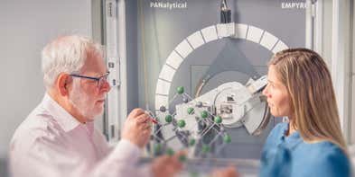

A non-destructive technique to study all types of material
In materials research, the scientist has many analytical questions related to the chemical composition and crystalline constitution of materials. X-ray diffraction (XRD) is the only laboratory technique that non-destructively and accurately obtains information such as chemical composition, crystal structure, crystal orientation, crystallite size, lattice strain, preferred orientation and layer thickness. Materials researchers therefore use XRD to analyze a wide range of materials, from powders to solids, thin films and nanomaterials.
X-ray diffraction (XRD) is a versatile non-destructive analytical technique used to analyze physical properties such as phase composition, crystal structure and orientation of powder, solid and liquid samples.
Many materials are made up of tiny crystallites. The chemical composition and structural type of these crystals is called their 'phase'. Materials can be single-phase or multiphase mixtures and may contain crystalline and non-crystalline components. In an X-ray diffractometer, different crystalline phases give different diffraction patterns. Phase identification can be performed by comparing X-ray diffraction patterns obtained from unknown samples to patterns in reference databases. This process is like matching fingerprints in a crime scene investigation.
The most comprehensive compound database is maintained by ICDD (International Center of Diffraction Data). You can also build a reference database from measured pure-phase diffraction patterns, from patterns published in the scientific literature, or from your own measurements. The relative strengths of the patterns from different phases in a multiphase mixture are used to determine the full composition of a sample.
An X-ray instrument contains three main items: an X-ray source, a sample holder and an XRD detector.
The X-rays produced by the source illuminate the sample. It is then diffracted by the sample phase and enters the detector. By moving the tube or sample and detector to change the diffraction angle (2θ, the angle between the incident and diffracted beams), the intensity is measured, and diffraction data are recorded. Depending on the geometry of the diffractometer and the type of sample, the angle between the incident beam and the sample can be either fixed or variable and is usually paired with the diffracted beam angle.
Many researchers, in industrial as well as in scientific laboratories, rely on X-ray diffraction (XRD) as a tool to develop new materials or to improve production efficiency. Innovations in X-ray diffraction closely follow the research on new materials, such as in semiconductor technologies or pharmaceutical investigations. Industrial research is directed toward the ever-increasing speed and efficiency of production processes. Fully automated X-ray diffraction analyses in mining and building materials production sites result in more cost-effective solutions for production control.
The main uses of X-ray diffraction are:
Qualitative and quantitative phase analysis of pure substances and mixtures. The most common method for phase analysis is often called 'X-ray powder diffraction' (XRPD).
Other X-ray diffraction techniques for materials that are not polycrystalline (for example single crystal semiconductor wafers or epitaxial layers) include high-resolution analysis of heteroepitaxial layers (HR-XRD). The analysis of these make use of Bragg’s Law, dynamical diffraction theory, and single crystal orientation, for both wafer as well as ingots.
Other methods that study the non-crystalline components of a material using various X-ray scattering methods, include Grazing incidence small-angle X-ray scattering (GISAXS), Small-angle X-ray scattering (SAXS), Total scattering (also called Pair Distribution Function (PDF) analysis), X-ray reflectometry (XRR). Each method has its own algorithm for data analysis, based on fundamental scattering theory.
Once an X-ray scattering or diffraction pattern has been measured. It needs to be analyzed. Analysis of X-ray diffraction and X-ray scattering data can be very complex. To make this easier for the user, a variety of XRD software packages exist to support all the different types of measurement.
XRD is rather fast (typically below 20 minutes) and is often the most accurate and reliable technique for the unambiguous identification of unknown materials.
Sample preparation is minimal which is a reason why this technique is so popular and is suitable for use both in industrial process applications and in materials research.
With the right analytical software, data analysis can be quite straightforward and for industrial processes, it can be even automated so that in QC applications the operator does not need to be an XRD expert.

Empyrean rangeMultipurpose X-ray diffractometers for your analytical needs |

AerisThe future is compact |
|
|---|---|---|
| Measurement type | ||
| Particle shape | ||
| Particle size | ||
| Crystal structure determination | ||
| Phase identification | ||
| Phase quantification | ||
| Contaminant detection and analysis | ||
| Epitaxy analysis | ||
| Interface roughness | ||
| 3D structure / imaging | ||
| Technology | ||
| X-ray Diffraction (XRD) | ||
| Goniometer configuration | Vertical goniometer, Θ-Θ and ω-Θ | Vertical goniometer, coupled and decoupled θ-θ, samples always horizontal |
| Detector | PIXcel1D, PIXcel3D, PIXcel3D 2x2, GaliPIX3D, Proportional counter, Scintilation detector | PIXcel1D, PIXcel3D and 1Der detectors |
| X-ray tube anode material | Cu, Co,Cr, Mn, Fe, Mo, Ag | Cu /Co (option) |