It is specifically stated in USP 788 that particulate matter consists of mobile undissolved particles, other than gas bubbles, unintentionally present in the solutions. For this and other USP chapters, particulate counts are required and it is preferable, although not required, to be able to indicate whether the particles counted are of interest, such as protein aggregates or large API particles in inhaled products, or simply excipients, silicone droplets or other contaminant of little health concern.
This application note describes counting particles on a filter membrane by automated particle imaging and discusses a means of validating this particle counting method.
1 ml of a 2000 particles/ml counting standard (Thermo Scientific Ezy-cal lot 37088) was filtered using a nitro-cellulose filter membrane with 0.45 µm pores. The nominal particle size is 10 μm and is certified to have 2000 (± 10%) particles per ml > 7.5 μm.
The Morphologi G3 was used to characterize the particle size and shape distributions as well as the particle count. The samples were analyzed using the 10X objective, which covers a nominal particle size range of 3.5 to 210 μm. To match the certification of the standard, all particles with a Circle Equivalent (CE) diameter < 7.5 μm were removed from the results using a software filter. The focus position was set equal to the filter surface and Z stacking was used to account for any warping of the filter.
Two separate filtrations of 1 ml of sample were prepared and each filter was analyzed 4 times to assess the instrument repeatability.
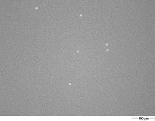
Figure 1 shows an example of a field of view captured during image acquisition.
The glass spheres are isolated from the filter membrane background based on the light intensity reflected to the camera with episcopic illumination.
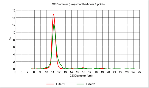
Figure 2 shows the size distribution of the population measured on the filters for the two samples. As expected, the size distributions overlay. Both distributions display small numbers of particles at ~16 and ~19 μm. These correspond to doublet and triplet particles.

|
Due to the particle doublets and triplets, a raw particle count will not give the correct number of spheres on the filter since counting multiple particles as one particle will lead to an underestimation of the number of spheres present. A classification scheme was therefore used to place the particles into classes according to the number of spheres present. The particles were separated based on CE diameter into singlets (< 15.1 µm), doublets (15.1 to 18 µm), triplets (18 to 21 µm), and multiplets (> 21 µm).
Quantifying the number of particles in each class allows for a more accurate determination of the number of spheres present than a raw particle count. Classifying particles allows for a doublet particle to be counted as

|
two spheres, a triplet as three spheres, and so on. This will lead to a much more accurate count that the raw particle count.
Table 1 shows the total number of particles detected along with the number of particles in each class for each repetition of the measurement of both filters. The last column indicates the number of individual spheres assuming the particles in the multiplets class contain an average of 5 spheres.
| Sample | Total Particles | Singlets | Doublets | Triplets | Multiplets | Spheres |
|---|---|---|---|---|---|---|
| 1940 | 1896 | 27 | 14 | 3 | 2007 | |
| 1 | 1958 | 1917 | 26 | 14 | 1 | 2016 |
| 1958 | 1914 | 27 | 14 | 3 | 2025 | |
| 1964 | 1917 | 32 | 14 | 1 | 2028 | |
| AV | 1955 | 2019 | ||||
| RSD % | 0.53 | 0.47 | ||||
| 1914 | 1840 | 30 | 28 | 16 | 2064 | |
| 2 | 1923 | 1845 | 34 | 30 | 14 | 2073 |
| 1929 | 1849 | 41 | 27 | 12 | 2072 | |
| 1914 | 1850 | 23 | 31 | 10 | 2039 | |
| AV | 1920 | 2062 | ||||
| RSD % | 0.38 | 0.77 |
The certified particle count was 2000 ± 10% per ml, therefore the expected number of particles is 2000 ± 200 for each measurement. For both samples the particle count is within the expected count range and in both cases, the relative standard deviation of the repeated measurement of each sample is less than 1%.
The Morphologi G3 was successful in counting the number of spheres collected on a filter from a particle counting standard. In each case, the resulting particle count was within the expectations based on the certified particle count in the standard. Additionally, the relative standard deviation in the number spheres detected for repeated measurements on the same filter was less than 1% indicating excellent repeatability.