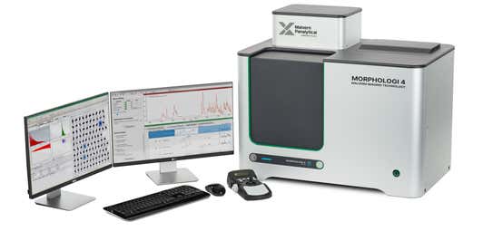

Automated direct measurement of particle size and particle shape
Morphological imaging applies the technique of automated static image analysis to provide a complete, detailed description of the morphological properties of particulate materials.
By combining particle size measurements, such as length and width, with particle shape assessments, such as circularity and convexity, morphological imaging fully characterizes both spherical and irregularly shaped particles. This enables a deeper understanding of a sample’s characteristics through precise detection of agglomerates, foreign particles and other anomalous materials. It also delivers the data required to cross-validate other particle sizing methods which apply an equivalent-sphere approach to reporting particle size distributions.
Morphological imaging precisely characterizes individual particles within pre-dispersed samples of dry powders, wet suspensions, and particulates deposited on filters. Statistically representative distributions are constructed by rapidly and automatically analyzing hundreds of thousands of particles per measurement, providing valuable information on the whole sample.


Combining static imaging with Raman spectroscopy in a technique known as Morphologically-directed Raman spectroscopy (MDRS) further allows the chemical identity of the particles to be determined, delivering component-specific microstructural insight. Raman spectroscopy is well-recognized across industry and academia as providing the high level of chemical specificity required to identify components in a mixture, even to the degree of differentiating alternative forms of the same compound.
Particulate samples can be prone to agglomeration which may not be easily detected by other particle sizing techniques. Analysis of individual particles in the dispersion in terms of their outline shape enables you to determine if, and to what extent, agglomerates are present.
Milling can change particle shape and size, which affects a material’s processing behavior and final properties. By measuring shape parameters such as elongation or circularity, the overall sample form is monitored and process changes can be made if required.
Powder flow and abrasive powder effectiveness are both influenced by particle surface texture. Particle shape parameters help assess if a powder is likely to stick in a hopper or if an abrasive powder has become worn.
Mineral samples often contain a mixture of different particle types. Using greyscale images to measure physical properties such as the amount of light passing through or being reflected from the particle’s surface helps to differentiate between these particles.

Morphologi rangeAutomated imaging for advanced particle characterization |
|
|---|---|
| Technology | |
| Image analysis | |
| Measurement type | |
| Particle shape | |
| Particle size | |
| Particle size range | 0.5µm - 1300µm |